Section 1: Product Introduction & Clinical Purpose
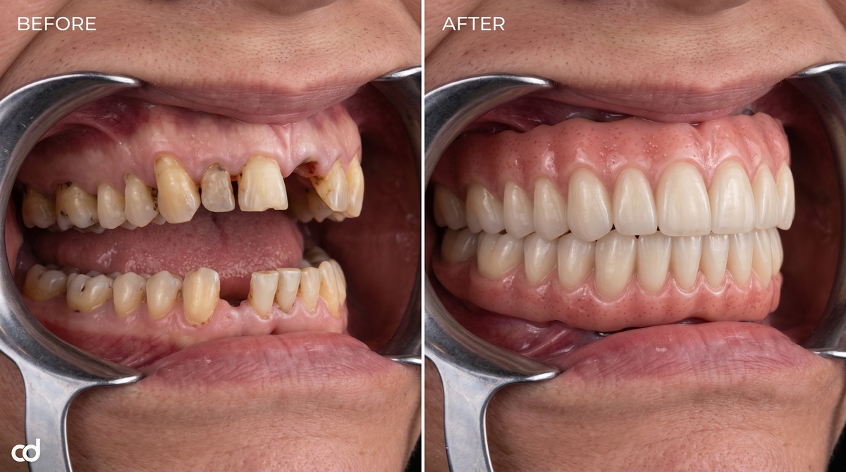
The Curedent Stackable Surgery Guide is an engineered, multi-tier CAD/CAM appliance designed to guide full-arch terminal dentition and edentulous reconstructions from initial osteotomy to chairside immediate loading. Built specifically for implant surgeons, periodontists, and prosthodontists performing full-arch rehabilitations, this system eliminates the clinical uncertainty of analog bone reduction, freehand pin placement, and compromised prosthetic conversions.
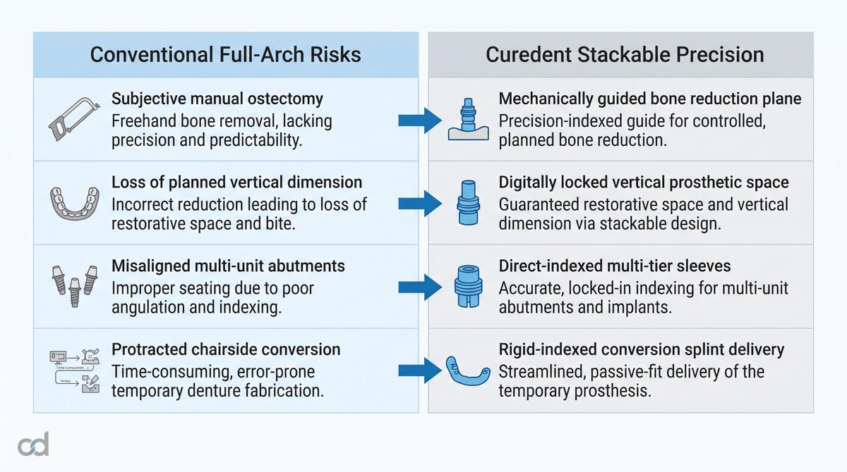
Executing All-on-X immediate function demands strict coordination between surgical ostectomy, precise implant trajectories, and prosthetic delivery within a single clinical appointment. In conventional freehand surgery, uneven manual bone flattening often compromises the prosthetic envelope, alters the vertical dimension of occlusion (VDO), and yields misaligned multi-unit abutments that complicate immediate conversion. The Curedent stackable system establishes a rigid, sequentially locked workflow where every surgical step indexes directly off an initial bone-anchored base.
Key Clinical Advantages
- Enforces Engineered Ostectomy: Establishes a predictable bone shelf that guarantees adequate space for the definitive prosthesis.
- Maintains Vertical Control: Locks the planned restorative vertical dimension across all surgical and conversion tiers.
- Eliminates Angular Guesswork: Mechanically controls multi-unit abutment pathways to ensure screw-retained accessibility.
- Accelerates Operatory Conversion: Integrates an indexed prosthetic splint to transition smoothly from implant torque validation to immediate provisionalization.
- Minimizes Operative Stress: Reduces intraoperative decision-making through a pre-planned, step-by-step modular surgical kit.
Section 2: Product Anatomy & Physical Construction
Medical-Grade Additive Polymer Matrix
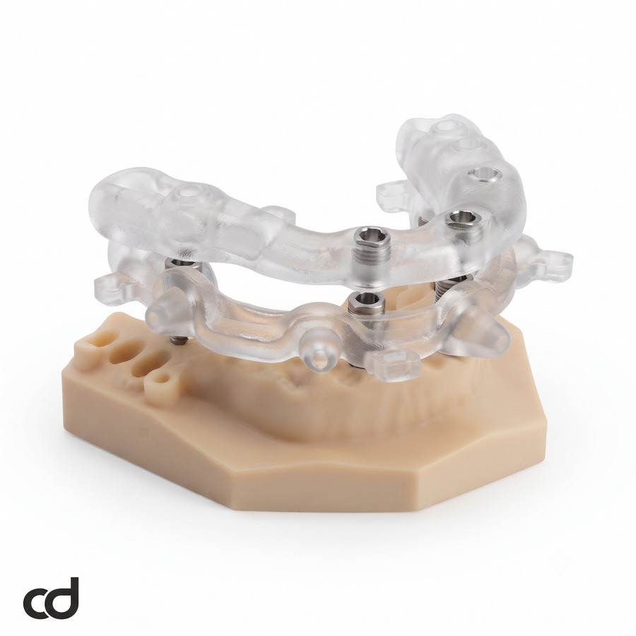
The Curedent Stackable Surgery Guide is fabricated from an FDA-compliant, Class IIa certified biocompatible photopolymer resin. This material is selected for its high flexural modulus, low water sorption, and resistance to torsional deformation under high surgical handpiece loads. The material’s optical transparency allows continuous visualization of the underlying osseous topography, cortical margins, and soft-tissue boundaries throughout surgical execution.
High-Resolution 3D Printing Architecture
Manufactured via high-precision Digital Light Processing (DLP) and Stereolithography (SLA) platforms at ~50-micron layer resolution, the guide components feature micro-accurate mating surfaces. This additive resolution captures minute geometric details, ensuring that the modular tiers dock without frictional binding, mechanical rocking, or linear seat discrepancy.
The Stackable Surgical Guide System Architecture
The stackable surgical guide system uses a modular, three-tier interlocking design:

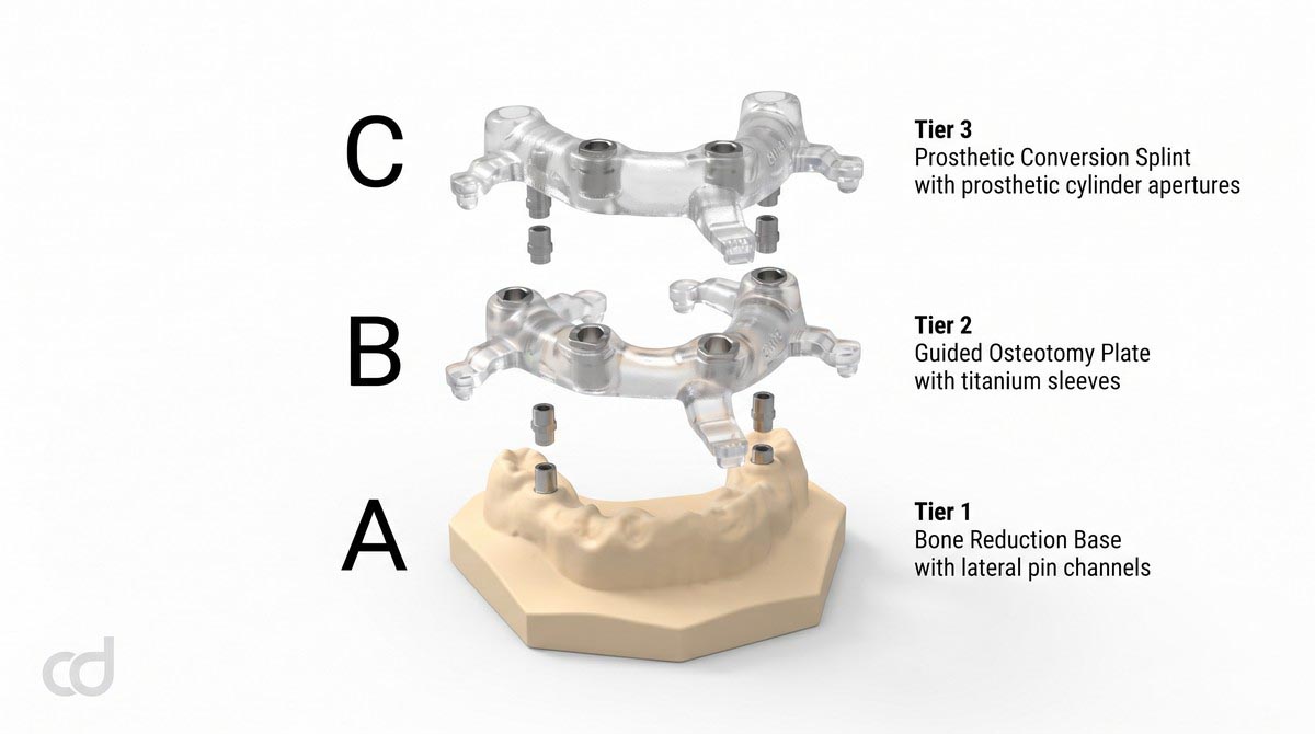
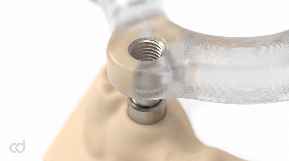
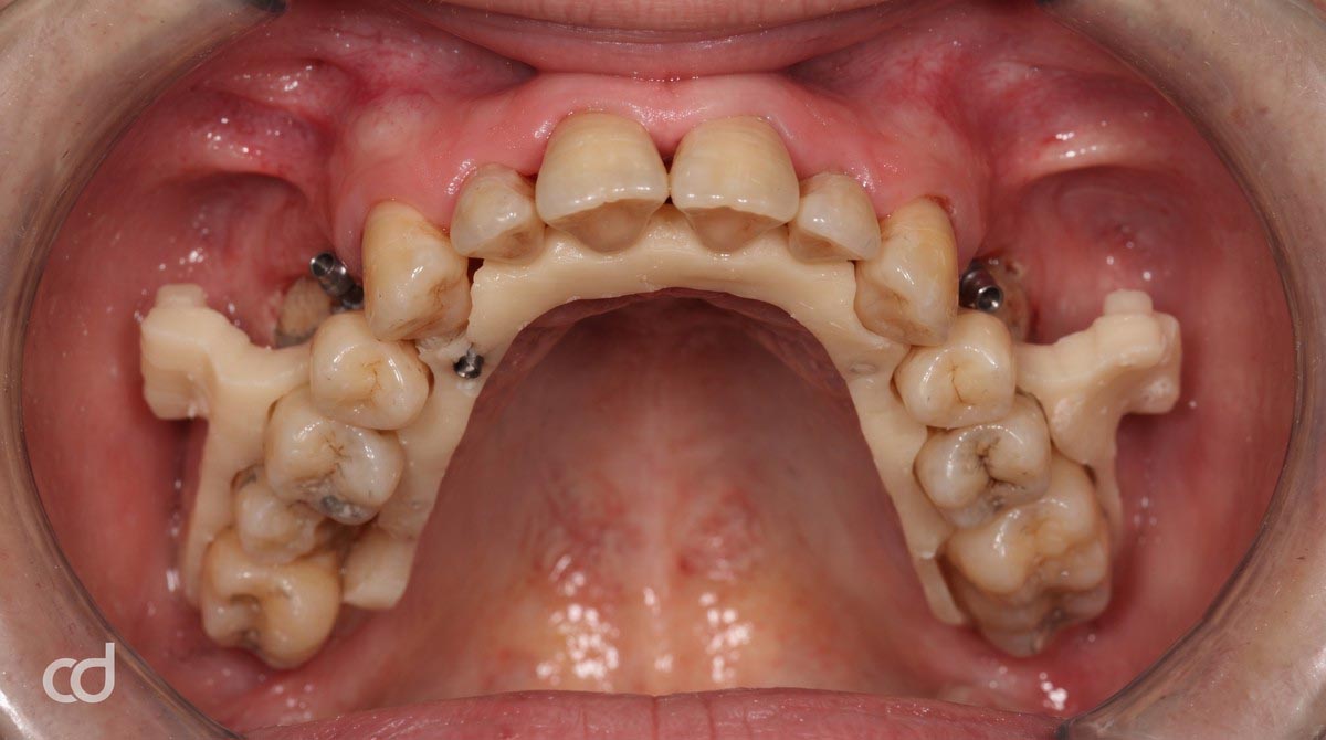
- Tier 1 (Fixation & Bone Reduction Base): Serves as the primary surgical foundation. It rests directly on intact anatomical landmarks (either remaining teeth via an initial seating template or cortical bone contours) and is locked in place using transverse anchor pins. Its coronal surface defines the precise bone reduction plane.
- Tier 2 (Osteotomy Guidance Template): Docks directly onto Tier 1 using high-tolerance mechanical interlocks. It houses calibrated medical-grade titanium master sleeves that guide pilot, shaping, and final osteotomy drills, as well as guided implant drivers.
- Tier 3 (Prosthetic Conversion Splint): Docks onto the anchored Tier 1 base following fixture placement. It guides temporary cylinder positioning, confirms multi-unit abutment orientation, and supports rapid pickup of the provisional immediate-load prosthesis.
Precision Indexing and Guide Rocking Prevention
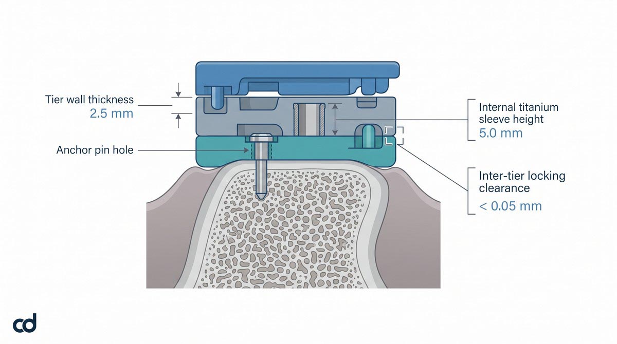
Mechanical stability across multi-tier surgical guides depends on positive indexing between components. The Curedent system incorporates cross-arch male-female locating pins, magnetic latches, and mechanical friction locks engineered to tolerances below 0.05 mm.
This positive registration ensures reliable guide rocking prevention, neutralizing rotational torque generated by high-speed surgical drills and bone saws. Once the surgeon seats an upper tier onto the Tier 1 base, tactile feedback confirms the component is locked, preventing axial displacement during surgery.
Seamless Chairside Tier Transitions
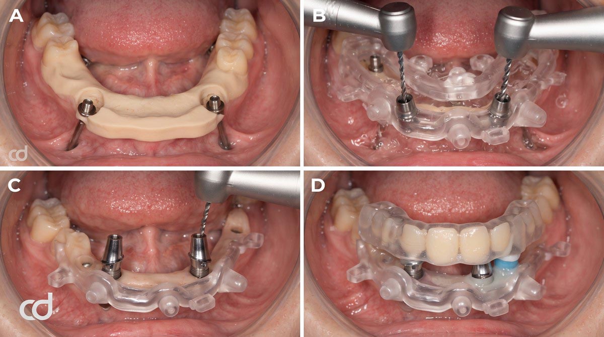
Tier transitions are designed for efficiency. Tier 1 remains rigidly pinned to the cortical bone throughout the entire surgery. Once bone reduction is complete along the Tier 1 platform, the surgeon seats Tier 2 onto Tier 1 without removing bone pins or disrupting surgical reference points. After implant insertion, Tier 2 is removed, and Tier 3 is engaged onto the same Tier 1 base to guide immediate prosthetic conversion.
Section 3: Indications & Clinical Applications
Primary Clinical Indications
The Curedent Stackable Surgery Guide is indicated for complex full-arch oral rehabilitations:
- Terminal Dentition Requiring Immediate Extraction and Full-Arch Rehabilitation: Cases with failing, broken, or periodontally involved teeth requiring full clearance, alveolectomy, and immediate implant placement.
- Edentulous Ridges with Irregular Osseous Topography: Patients with knife-edge ridges, severe vertical bone discrepancies, or exostoses requiring uniform bone flattening to establish a level restorative base.
- All-on-4, All-on-5, and All-on-6 Immediate-Load Workflows: Full-arch protocols where simultaneous fixture placement, multi-unit abutment alignment, and immediate provisionalization are performed within a single visit.
- Cases Requiring Controlled Bone Reduction: Procedures where prosthetic clearance must be carefully balanced against available bone volume to avoid compromising anatomic structures.
Full-Arch Diagnostic Checkpoints:
[✓] Diagnostic smile design confirms lip-line transition zone
[✓] Restorative space calculation: >= 15 mm from bone plane to incisal edge
[✓] Sufficient basal bone volume apical to the reduction plane
[✓] Minimum 3 transverse anchor pin sites with intact buccal-lingual cortical plates
[✗] Insufficient vertical bone volume where ostectomy compromises implant stability
Case Selection Criteria
- Adequate Basal Bone Volume: Following digital bone reduction, there must remain sufficient bone height and width (>10 mm height, >6 mm width) to achieve primary implant stability (35 Ncm).
- Keratinized Tissue Dimensions: Favorable bands of keratinized tissue must be available to allow mucosal management around prosthetic cylinders and prevent chronic peri-implant inflammation.
- Adequate Maxillomandibular Opening: Patients must have sufficient vertical opening (40 mm) to accommodate long drill barrels, reduction keys, and stackable guide components in posterior sectors.
The Biological Restorative Space (15mm) Principle
A common failure mode in full-arch fixed implant prosthetics is prosthetic fracture or aesthetic compromise resulting from inadequate restorative height. The transition between the patient’s natural tissue and the prosthetic pink acrylic must remain hidden apical to the dynamic high smile line.
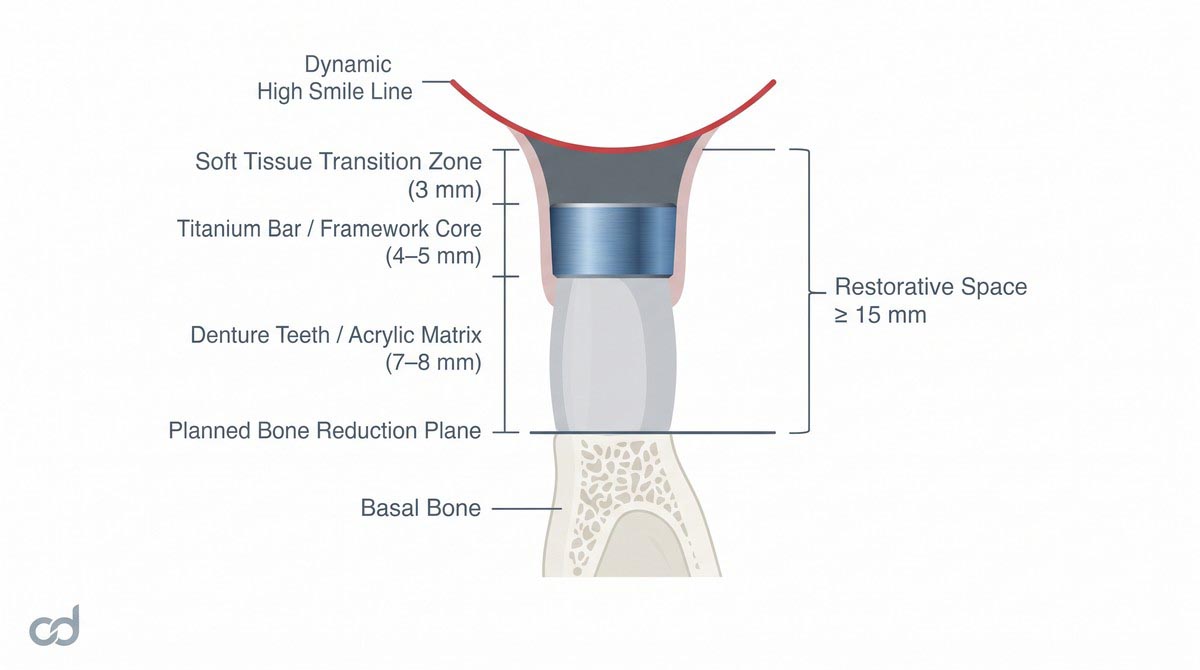
Achieving this transition requires establishing adequate biological restorative space (15mm) measured from the prepared bone crest to the planned incisal edge. This 15 mm clearance accommodates:
- Space for the multi-unit abutment and prosthetic cylinder (3–4 mm).
- Adequate bulk of high-strength structural framework (4–5 mm).
- Sufficient thickness of restorative acrylic and denture teeth (7–8 mm) to resist functional shear and fracture.
Defining the Bone Reduction Plane
The Curedent digital workflow maps this vertical requirement directly into the bone reduction plane. During virtual planning, the software simulates extractions, aligns the patient’s high smile line, and sets a horizontal ostectomy shelf that guarantees 15 mm of restorative space while preserving safe clearance above the inferior alveolar nerve canals or beneath the maxillary sinus floors. Tier 1 physically materializes this virtual plane at the chairside.
Section 4: Clinical Workflow & Surgical Execution
Step 1: Comprehensive Data Acquisition
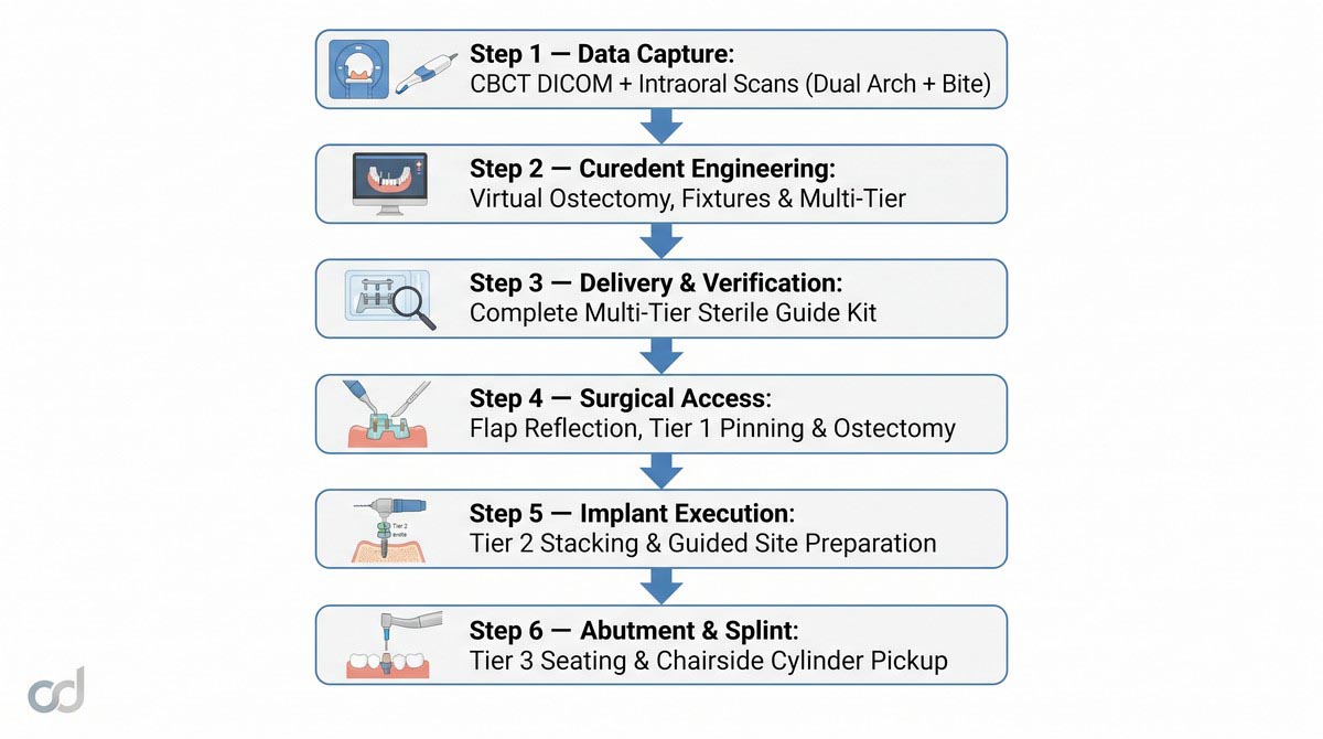
Acquire a high-resolution CBCT DICOM dataset of the affected arch with the patient in maximum intercuspation, ensuring the mandibular canal and maxillary sinuses are completely captured without patient motion artifacts. Perform optical intraoral scans of the maxillary and mandibular arches, recording accurate soft-tissue contours, palate anatomy, and interocclusal records.
Step 2: Virtual Digital Engineering by Curedent
Upload the DICOM and optical STL files to the Curedent portal. Curedent engineers register the digital models, integrate the diagnostic tooth setup, determine the ostectomy shelf to secure the 15 mm biological space, plan implant trajectories, optimize multi-unit abutment angles, and design the three-tier stackable system. The surgical team reviews and approves the virtual plan.
Step 3: Unboxing and Surgical Preparation
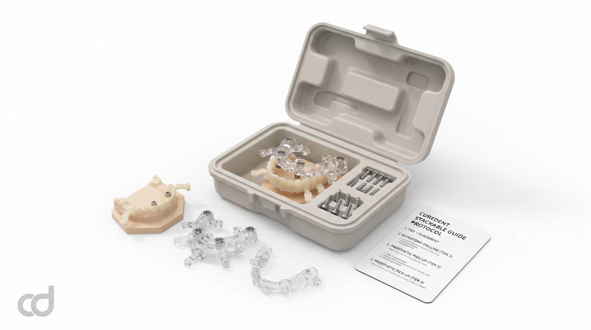
The guide system arrives in a dedicated, sterilization-ready package. Before entering the operatory, the team inspects the kit to confirm the presence of:
- Tier 1 Fixation & Bone Reduction Base
- Tier 2 Guided Osteotomy Template with embedded titanium sleeves
- Tier 3 Prosthetic Conversion Splint
- Intermediate tooth-supported positioning guide (for dentate cases)
- Calibrated horizontal anchor pins and dedicated drill keys
- Surgical plan printout indicating sleeve offsets, drill lengths, and angulations
Step 4: Surgical Flap Reflection, Tier 1 Pinning, and Ostectomy
- Administer local anesthesia. Extract non-retained teeth required for guide clearance while preserving any designated reference anchor units.
- Elevate a full-thickness mucoperiosteal flap past the mucogingival junction, exposing the facial and lingual cortical plates along the planned osteotomy line.
- Seat the Tier 1 guide using the tooth-supported positioning template (or bone-borne indexing pads in fully edentulous ridges).
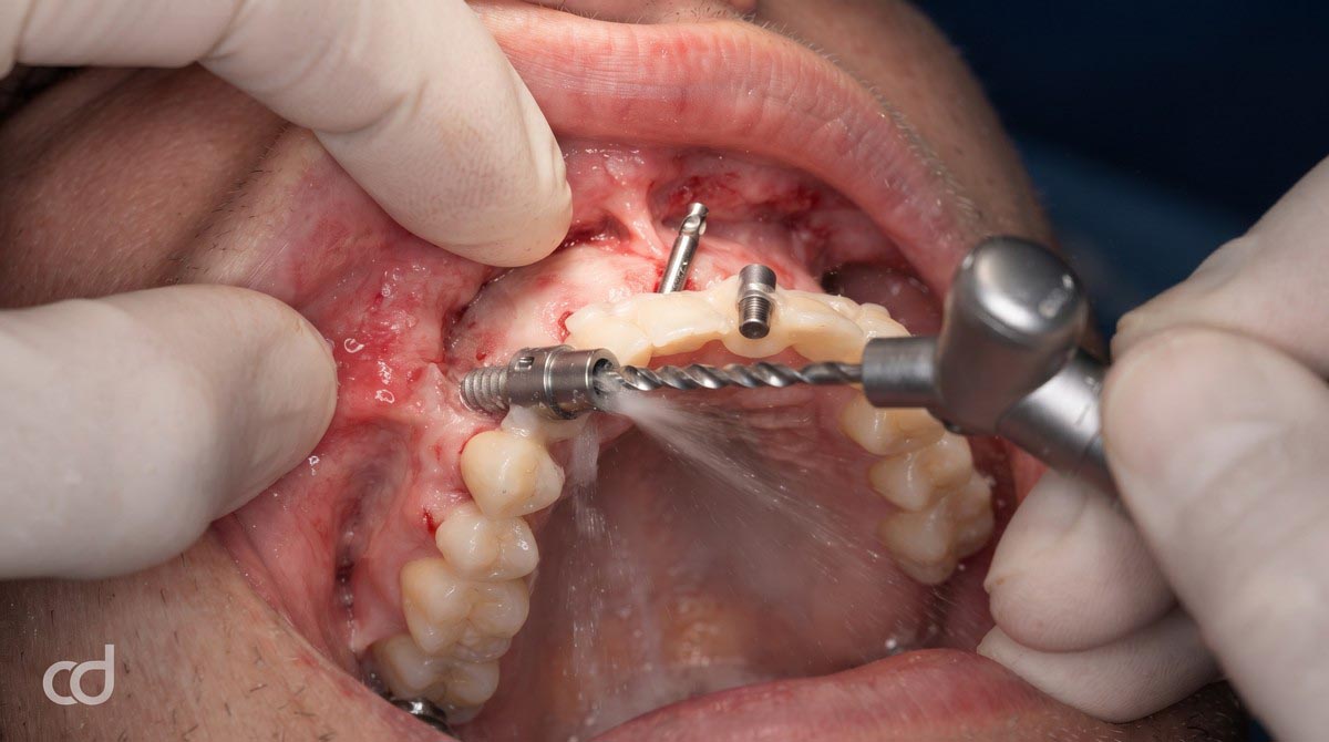
- Drill anchor pin channels through the guide ports using the dedicated pin drill. Ensure continuous irrigation.
- Drive the anchor pins to secure cortical bone engagement. The cross-arch triangulation of these transverse pins provides high anchor pin stability, establishing an immovable foundation that resists operative forces.
- Remove the preliminary positioning template, leaving Tier 1 firmly pinned.
- Perform the ostectomy using a surgical reciprocating saw or piezoelectric blade, gliding the cutting tool flush along the superior metal-reinforced shelf of Tier 1 until the entire arch matches the planned reduction plane.

Step 5: Tier 2 Stacking and Guided Osteotomy Preparation
- Snap Tier 2 directly onto the pinned Tier 1 base. Confirm complete mechanical seating across all cross-arch indexing junctions.
- Insert system-specific reduction keys into the titanium master sleeves.
- Advance the osteotomy drill sequence under continuous, copious saline irrigation. The sleeves guide drill path and depth, locking trajectory across anterior straight and posterior tilted sites.
- Insert implant fixtures directly through the Tier 2 sleeves using guided drivers until the mechanical stop collar contacts the sleeve rim. Verify primary stability (35 Ncm).
Step 6: Abutment Delivery and Tier 3 Prosthetic Conversion
- Remove Tier 2 from the pinned Tier 1 foundation.
- Seat designated multi-unit abutments onto the implants according to the surgical protocol plan. Torque to the manufacturer’s specified tension (15–35 Ncm).
- Place temporary titanium cylinders onto the multi-unit abutments.
- Snap Tier 3 (Prosthetic Conversion Splint) onto the Tier 1 base. The splint’s oversized access chimneys seat passively over the cylinders without binding.
- Apply dual-cure composite or PMMA pickup material through the chimneys to lute the cylinders to the splint.
- After polymerization, unthread the prosthetic retaining screws, release Tier 3, unpin and remove Tier 1, and transfer the conversion prosthesis to the laboratory bench for finishing and polishing.
Anchor Pin Stability and Soft-Tissue Management
Careful soft-tissue management prevents complications during fixation. The mucoperiosteal flap must be adequately released and retracted beyond the pin entry ports. Driving pins through unreflected mucosal margins can drag soft tissue into the bone channel, causing pin toggle and localized necrosis. Pin drills should engage both facial and lingual cortical plates to resist lateral lever forces without stripping the bone threads.
Section 5: Technical Specifications & Accuracy Standards
Certified Specification Standards
The Curedent Stackable Surgery Guide system is manufactured to tight mechanical tolerances, ensuring seamless tier interchangeability and reliable clinical transfer.
| Parameter | Specification | Clinical Relevance |
| Material | Class IIa biocompatible surgical resin | High torsional rigidity, zero intraoral toxic leaching |
| Printing method | DLP / SLA stereolithography | Captures micro-indexing interlocks without distortion |
| Layer resolution | ~50 microns | Ensures tight inter-tier fit and passive seating |
| Guide architecture | Multi-tier stackable (Tier 1 / Tier 2 / Tier 3) | Modular progression across reduction, osteotomy, and pickup |
| Anchor pin hole tolerance | 15–30 microns | Prevents anchor pin toggle and lateral base movement |
| Sleeve tolerance | 15–30 microns | Minimizes drill wobble while preventing thermal binding |
| Guide type | Bone-supported / pin-anchored full-arch system | Delivers cross-arch stability independent of remaining teeth |
| Tier-to-tier fit tolerance | < 0.05 mm | Eliminates cumulative vertical and angular tracking errors |
| Multi-unit abutment orientation accuracy | < 2° deviation | Ensures prosthetic screw access emerges through planned cingula |
| Sterilization | Autoclavable / sterilization-ready delivery | Complies with sterile surgical operatory protocols |
Accuracy Metrics and Cumulative Error Control
Full-arch guided procedures carry the risk of cumulative error propagation: minor shifts at the anchor pins can amplify into significant displacement at the restorative platform. The Curedent stackable system controls this through strict manufacturing metrics:

- Multi-Unit Abutment Orientation: Digital alignment presets the rotational timing and trajectory of angled multi-unit abutments (17,30). Curedent maintains multi-unit abutment orientation to within <2 of planned trajectory, ensuring that screw access holes emerge through the occlusal troughs or lingual cingula rather than the facial aesthetic zone.
- Inter-Tier Tolerances: By holding tier-to-tier interface tolerances below 0.05 mm, the system prevents vertical creep, ensuring that the bone reduction plane, implant depths, and conversion splint remain locked to the same spatial coordinates.
- Rigid Pin Stability: Anchor pin sleeves maintain tolerances within 15–30 microns, eliminating lateral rotational drift during full-arch bone sawing and heavy handpiece instrumentation.
Section 6: Clinical Benefits & Practice Impact
Patient-Level Clinical Benefits
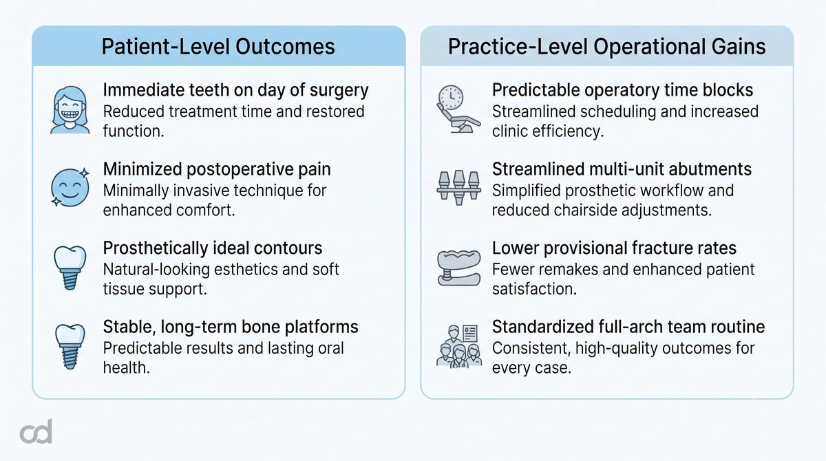
- Predictable Full-Arch Restorations: Patients receive a secure, rigid provisional restoration on the day of surgery, avoiding the discomfort and instability of traditional removable complete dentures.
- Reduced Postoperative Morbidity: Guided ostectomy and controlled flap elevation limit surgical trauma, minimizing postoperative swelling, ecchymosis, and pain.
- Optimized Emergence and Hygiene Access: Predictable bone reduction positions the prosthetic transition zone beneath the smile line while creating smooth, cleansable pontic contours that support long-term peri-implant health.
Practice-Level Operational Advantages
- Routine Adoption of Full-Arch Protocols: The stackable system removes the clinical uncertainty of freehand ostectomy and complex implant angulation, allowing surgical teams to adopt full-arch immediate-load treatments as a predictable clinical standard.
- Workflow Cost-Efficiency: Streamlining osteotomy, fixture placement, and provisional pickup within a unified system shortens surgical chair time, reduces consumable waste, and minimizes remakes of fractured conversion appliances.
- Reduced Intraoperative Stress: Pre-planned bone reduction and guided drilling eliminate real-time anatomical guesswork, allowing the team to focus on primary stability and soft-tissue closure.
- Simplified Laboratory Conversions: Using an indexed prosthetic conversion splint allows chairside assistants and laboratory technicians to lute temporary cylinders predictably, avoiding the extensive occlusion adjustments common in freehand conversion procedures.
The Standard for Immediate Full-Arch Care
Achieving predictable All-on-X immediate function requires shifting diagnostic and surgical decisions into the preoperative digital workflow. The stackable surgical guide system replaces analog freehand surgery with an integrated engineering process. By locking the bone reduction plane, osteotomy paths, and restorative conversion to an initial rigid foundation, clinicians can deliver immediate full-arch rehabilitations with high functional and aesthetic confidence.
Section 7: Ordering, FAQ & Call to Action
Simple Three-Step Ordering Workflow
Integrating the Curedent Stackable Surgery Guide into your full-arch workflow is designed to be straightforward:
- Upload Case Records: Access the Curedent portal and upload the patient’s CBCT DICOM files alongside optical intraoral scans (maxillary, mandibular, and dynamic interocclusal records).
- Interactive Digital Treatment Review: Collaborate online with Curedent biomedical engineers. Evaluate bone reduction levels, confirm implant positions and angulations, check restorative clearance, and approve the multi-tier guide configuration.
- Receive the Surgical Kit: Curedent fabricates the stackable guide set at ~50-micron resolution, confirms component fit, packages the assembly in protective, sterilization-ready casing, and dispatches the kit directly to your operatory.
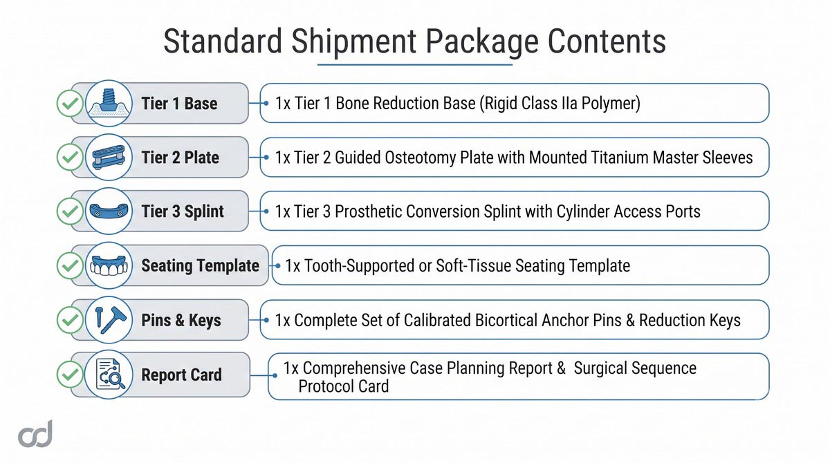
Standard Shipment Package Contents:
———————————–
[✓] 1x Tier 1 Bone Reduction Base (Rigid Class IIa Polymer)
[✓] 1x Tier 2 Guided Osteotomy Plate with Mounted Titanium Master Sleeves
[✓] 1x Tier 3 Prosthetic Conversion Splint with Cylinder Access Ports
[✓] 1x Tooth-Supported or Soft-Tissue Seating Template (For Initial Tier 1 Placement)
[✓] 1x Complete Set of Calibrated Bicortical Anchor Pins & Reduction Keys
[✓] 1x Comprehensive Case Planning Report & Surgical Sequence Protocol Card
Frequently Asked Questions
Is a CBCT scan required for every stackable guide case?
Yes. Accurate virtual planning requires high-resolution CBCT DICOM datasets to map cortical bone volume, determine nerve locations, calculate bone reduction levels, and ensure anchor pins achieve bicortical purchase.
Can I use my preferred implant surgical system with this guide?
Yes. Curedent titanium master sleeves are custom-calibrated to match all major commercial guided surgery kits. Simply specify your implant brand, fixture line, and guided instrumentation kit during portal upload.
How many tiers are included in the standard stackable system?
The standard package includes three functional tiers: Tier 1 (Bone Reduction Base), Tier 2 (Osteotomy Guidance Template), and Tier 3 (Prosthetic Conversion Splint), along with an initial indexing template to seat and pin Tier 1.
What corrective steps should be taken if Tier 1 does not seat passively?
If Tier 1 binds against bone during trial seating, inspect the lingual and facial cortical margins for unreflected periosteum or localized bone prominences. Lightly smooth minor bone high spots with a round bur until the base seats flush without rocking.
Is the Curedent 3D printing resin certified for intraoral surgical contact?
Yes. All guide components are 3D-printed using certified Class IIa biocompatible surgical resins that meet stringent international cytotoxicity, irritation, and systemic toxicity standards.
Suggested Clinical Solutions
- For single-tooth or short-span edentulous spaces, utilize the Curedent Single Implant Surgical Guide.
- Achieve precise aesthetic crown lengthening with the Curedent Gum Reduction Guide.
- Stabilize post-treatment occlusion and prevent nocturnal wear using Curedent Night Guards.