Section 1: Product Introduction & Clinical Purpose
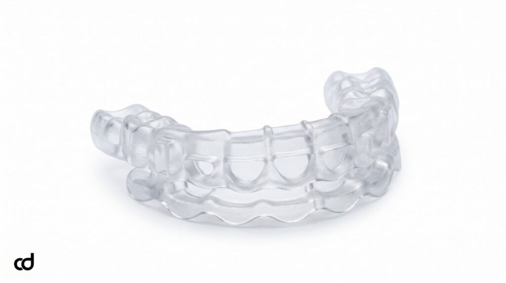
The Curedent Gum Reduction Guide is a patient-specific, CAD/CAM-manufactured surgical appliance engineered to guide both soft-tissue excision and crestal bone recontouring within a single clinical procedure. Built specifically for periodontists, aesthetic restorative dentists, and prosthodontists, this appliance translates anterior smile design diagnostics directly into operative predictability.
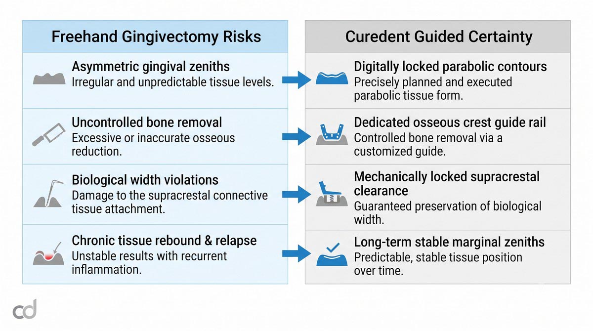
Freehand aesthetic crown lengthening often introduces intraoperative discrepancies. Tactile estimation frequently yields uneven scalloping, asymmetrical gingival zeniths across contralateral teeth, and uncalculated bone reduction that violates anatomical boundaries. Using the Curedent gingivectomy surgical guide, clinicians replace freehand guesswork with a mechanically locked template that transfers planned clinical crown heights and balanced gingival zeniths directly to the oral cavity.
Key Clinical Advantages
- Enforces Symmetrical Gingival Zeniths: Pre-indexes natural parabolic zeniths to balance central incisors, lateral incisors, and canines.
- Guarantees Biological Width Safety: Provides a physical stop for crestal ostectomy to prevent soft-tissue rebound and chronic inflammation.
- Dual-Tier Surgical Guidance: Integrates soft-tissue excision and hard-tissue recontouring into one stable tooth-borne template.
- Accelerates Restorative Timelines: Eliminates unexpected tissue rebound, enabling restorative preparation and final impressions without protracted corrective delays.
Section 2: Product Anatomy & Physical Construction
Medical-Grade Biocompatible Resin Matrix
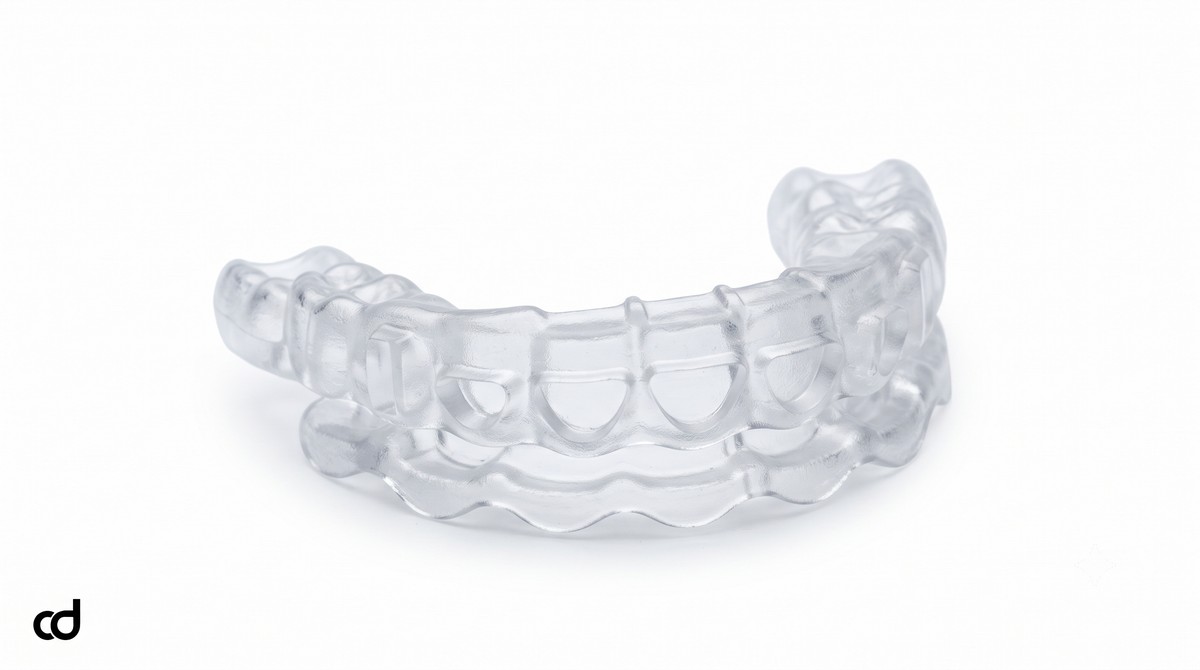
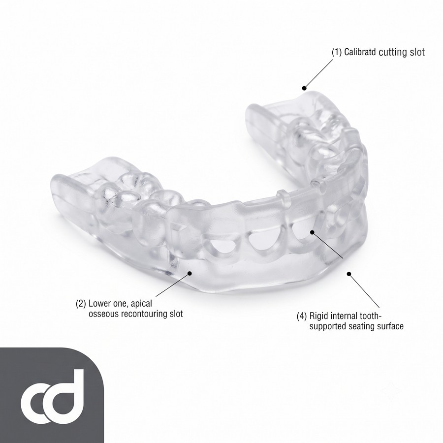
The Curedent gum reduction template is 3D-printed from an FDA-compliant, Class IIa certified biocompatible surgical polymer. This photopolymer is selected for its high dimensional stability, flexural strength, and optical transparency. The rigid polymer resists deformation during seating while providing clear visualization of the underlying gingival margin, tooth structure, and surgical cutting instruments.
High-Resolution Additive Manufacturing
Fabricated using industrial-grade Digital Light Processing (DLP) and Stereolithography (SLA) platforms at ~50-micron layer resolution, the guide captures fine occlusal morphology, contact points, and cervical embrasures. This fine layer resolution eliminates stepped edges within the cutting slots, ensuring scalpels, laser tips, or rotary burs travel smoothly along the guide paths without mechanical catch.
The Dual-Slot Gingival-Osseous Guide Architecture
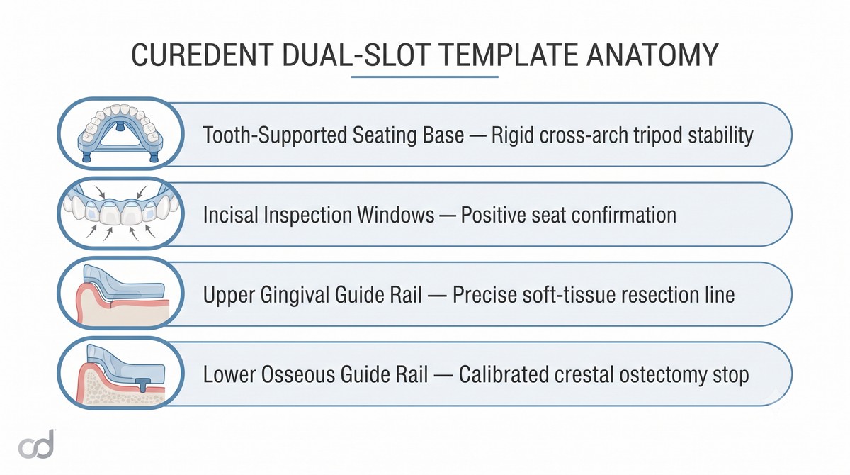
The core of the appliance is its dual-slot gingival-osseous guide framework. The template incorporates two distinct, parallel guidance rails for each tooth:
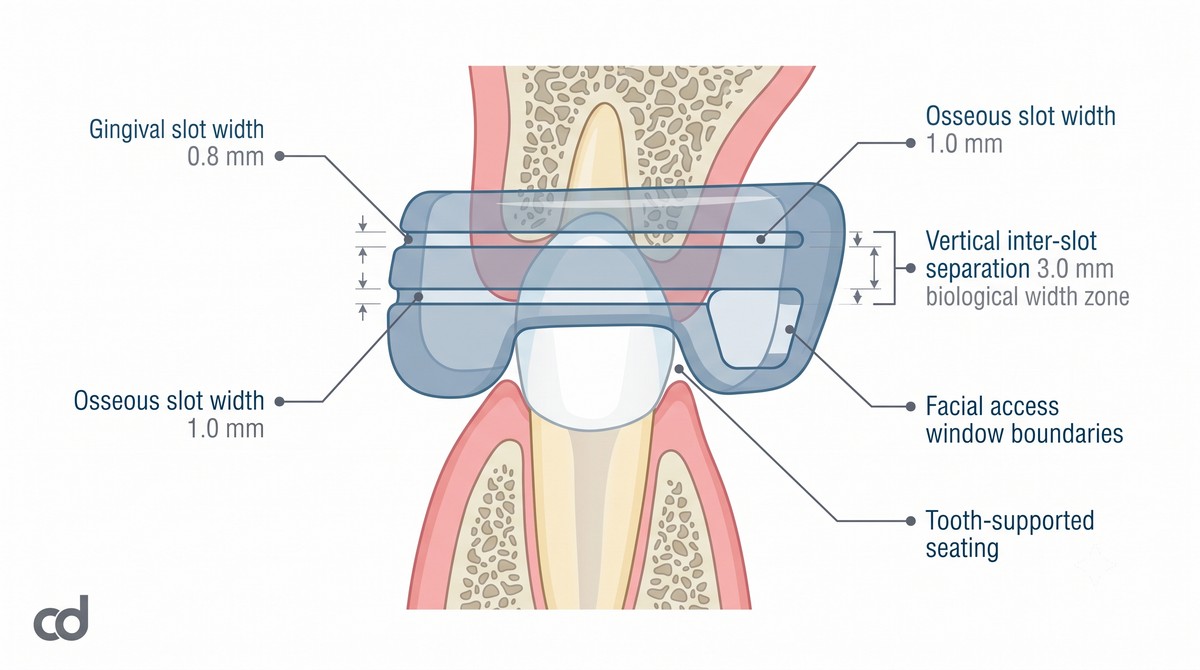
- Gingival Slot (Coronal Rail): Follows the planned parabolic free gingival margin, establishing the exact entry angle for surgical blades or laser tips during gingivectomy.
- Osseous Slot (Apical Rail): Positioned apically at an engineered offset distance, establishing the reference line for bone sounding and crestal ostectomy.
Windowed Structural Framework
Designed as a windowed surgical template, the appliance features open anterior apertures and incisal viewports. The open labial windows expose the exact surgical field, facilitating surgical access, continuous cold saline cooling, and visual monitoring, while the occlusal windows confirm complete seating before surgery begins.
Single-Position Dual-Tissue Management
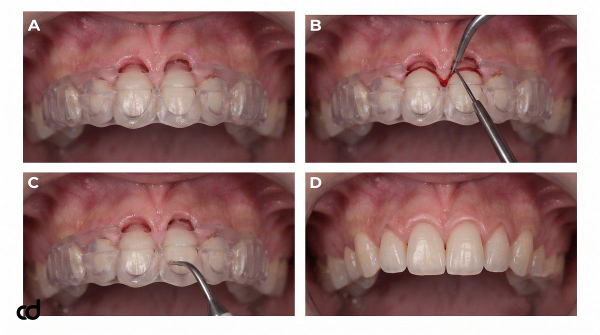
In conventional crown lengthening, flap reflection often disrupts visual landmarks, making it difficult to reference the original gingival margin during bone removal. The Curedent dual-slot design solves this problem by remaining fully seated throughout both soft-tissue resection and bone recontouring. The surgeon performs the initial gingivectomy through the coronal slot, reflects a conservative micro-flap through the labial window, and completes the ostectomy through the lower slot—all without altering the guide’s reference position.
Section 3: Indications & Clinical Applications
Primary Clinical Indications
The Curedent Gum Reduction Guide is indicated across several aesthetic anterior applications:
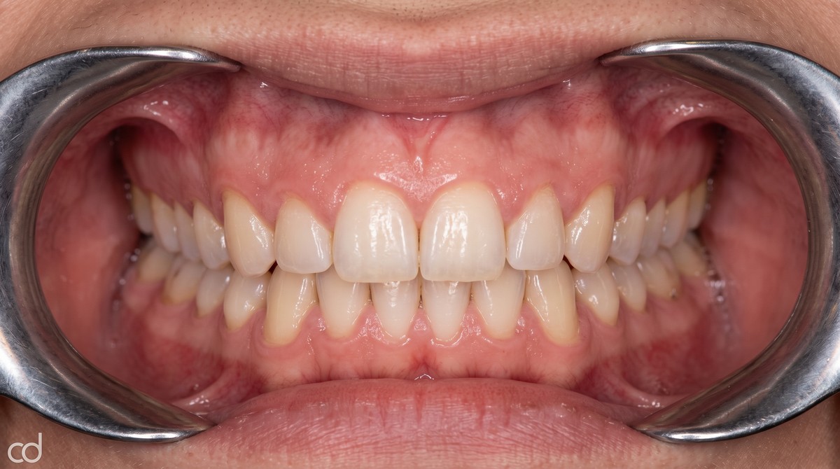
- Altered Passive Eruption (Type I and Type II): Clinical scenarios where the gingival complex failed to migrate apically during tooth eruption, leaving clinical crowns short and square with excessive gingival display.
- Gummy Smile (Excessive Gingival Display): Cases involving hypermobile upper lips or vertical maxillary discrepancies that require aesthetic recontouring to establish a balanced tooth-to-gingiva ratio.
- Asymmetric Gingival Margins: Discrepant gingival zeniths between central incisors, lateral incisors, and canines prior to restorative veneer or crown placement.
- Pre-Restorative Space Creation: Localized clinical crown lengthening required to establish ferrule and clear margins without violating anatomical attachment zones.
Aesthetic Crown Lengthening Feasibility Checklist:
[✓] Periodontal screening: Pocket depths stable, no active inflammation
[✓] Keratinized tissue: >= 2 mm of attached gingiva preserved post-resection
[✓] Crown-to-root ratio: Favorable post-surgical ratio (>= 1:1.5)
[✓] Alveolar crest location: Verified via transgingival bone sounding
[✗] Inadequate attached tissue leading to alveolar mucosal margin (Contraindicated)
Case Selection Criteria
- Adequate Keratinized Tissue Band: The patient must present with a sufficiently wide zone of keratinized gingiva so that at least 2.0 mm of attached gingiva remains after excision. Resecting beyond the mucogingival junction into alveolar mucosa is contraindicated.
- Stable Periodontal Health: The periodontium must be free from active plaque-induced gingivitis or periodontitis, with physiological probing depths (<3 mm) and healthy tissue architecture.
- Favorable Crown-to-Root Ratio: Anticipated osseous reduction must preserve adequate root anchorage, ensuring a postoperative crown-to-root ratio of at least 1:1.5.
Clinical Management of Altered Passive Eruption
In patients presenting with altered passive eruption, the excessive gingival drape hides the anatomical crown beneath the cementoenamel junction (CEJ). Using an aesthetic crown lengthening stent provides a reproducible method to expose the natural enamel contours. The guide maps the planned zenith slightly distal to the long axis on central incisors and canines, matching natural aesthetics.
Preserving Supracrestal Tissue Attachment (Biological Width)
A major cause of postoperative complications in aesthetic periodontal surgery is the violation of the supracrestal tissue attachment (biological width). The natural dentogingival complex requires a biological dimension—traditionally recognized as 2.04 mm, comprising approximately 0.97 mm of junctional epithelium and 1.07 mm of supracrestal connective tissue attachment—plus a 1.0 mm physiological sulcus depth.
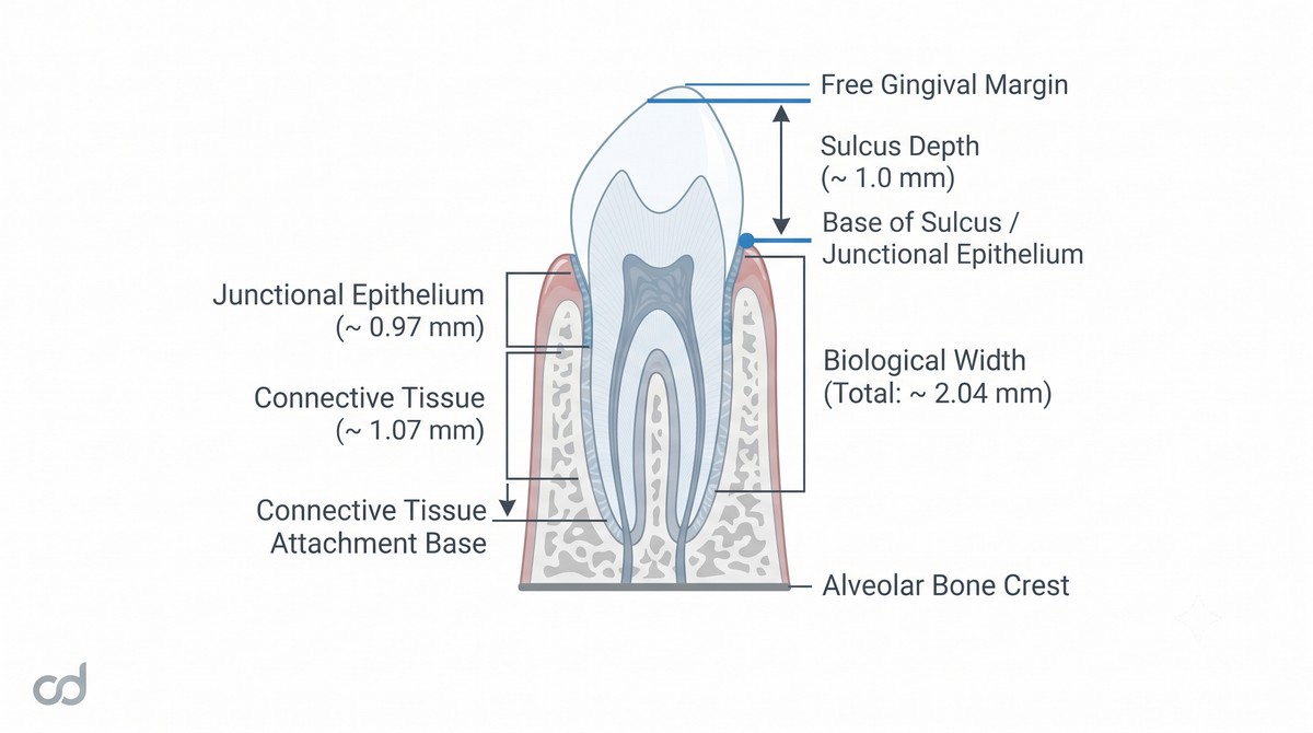
If a clinician resects gingival soft tissue without establishing at least 3.0 mm of clearance between the new gingival margin and the alveolar crest, the body re-establishes its biological seal through coronal tissue rebound, chronic marginal erythema, and unpredictable pocket formation. The Curedent appliance prevents this complication: the osseous slot is positioned precisely 3.0 mm apical to the gingival resection rail, mechanically ensuring that crestal bone reduction preserves the natural supracrestal attachment apparatus.
Section 4: Clinical Workflow & Surgical Execution
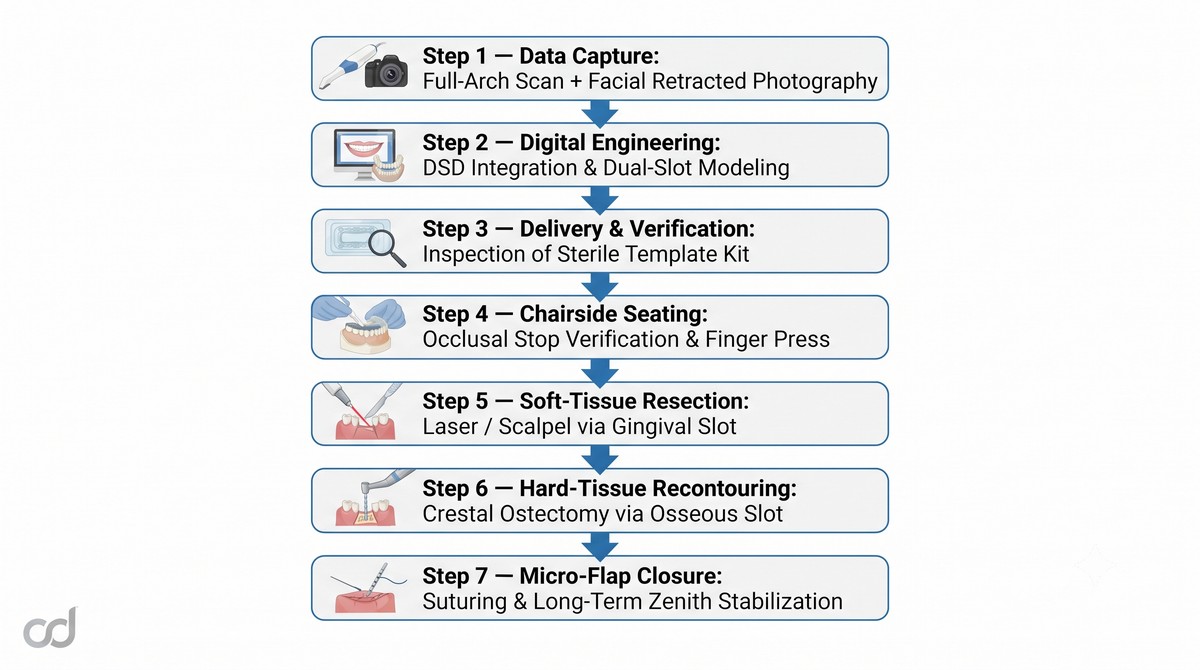
Step 1: Digital Data Acquisition
Capture high-resolution optical intraoral scans of the maxillary and mandibular arches, recording accurate soft-tissue topography and tooth margins. Acquire a standardized photographic series: 1:1 retracted frontal view, high dynamic smile view, and 45-degree aesthetic angles.
Step 2: Digital Planning and DSD Integration
Upload the intraoral STL files and photographic series to the Curedent portal. Through digital smile design (DSD) integration, Curedent engineers map the facial midline, interpupillary line, and dynamic lip contours to establish target tooth proportions (width-to-length ratio of 75–80%). The software calculates the ideal gingival zenith trajectory and models the dual-slot guide rails. The clinician reviews and approves the digital proposal.
Step 3: Unboxing and Pre-Surgical Verification
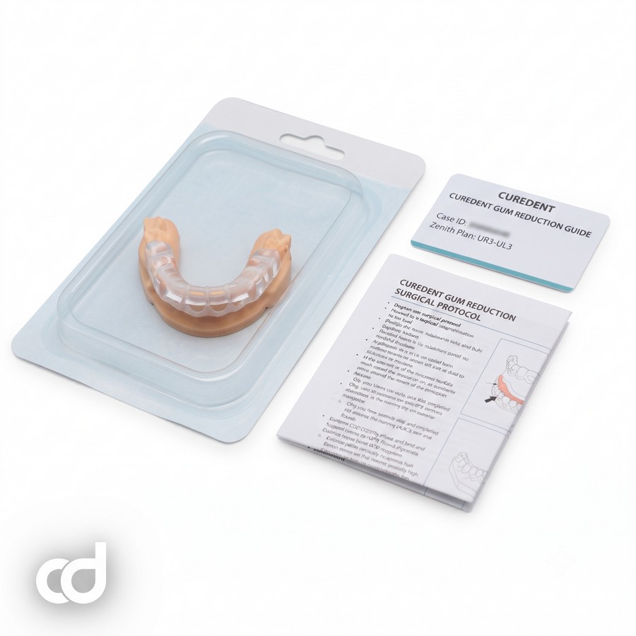
The guide arrives in dedicated, sterilization-ready packaging. Prior to surgery, confirm the patient name and site designation. Verify that the appliance is clean and free of micro-flashes, that the dual cutting slots are patent, and that the tooth-bearing retention base is structurally intact.
Step 4: Chairside Seating and Fit Verification
- Administer local infiltration anesthesia apical to the mucogingival junction to minimize soft-tissue distortion around the marginal gingiva.
- Seat the guide over the dentition. Apply firm, downward finger pressure along the incisal edges.
- Inspect every incisal inspection window to confirm complete, gap-free contact with the enamel.
- Confirm cross-arch stability, ensuring there is no rotational play or elastic rocking.
Step 5: Guided Gingival Resection
Engage surgical blade, micro-scalpel, or diode/erbium dental laser tip directly into the coronal gingival slot. Follow the engineered contour smoothly from distal line angle to mesial line angle. The guide rail directs the blade along the planned parabolic zenith while establishing an internal bevel angle of 45 degrees toward the tooth surface. Remove the excised marginal collar.

Step 6: Guided Crestal Ostectomy
- Elevate a conservative, full-thickness sulcular micro-flap through the wide labial window to expose the alveolar bone crest, leaving the guide fully seated.
- Introduce an end-cutting bur, micro-chisel, or piezoelectric bone tip into the apical osseous slot.
- Resect the bone crest flush with the lower guide rail under continuous saline cooling. This step re-establishes the 3.0 mm biological distance between the newly contoured gingival margin and the alveolar bone shelf.
- Perform delicate interproximal osteoplasty to create a smooth, scalloped osseous architecture that mirrors the planned soft-tissue profile.
Step 7: Final Irrigation and Hemostasis
Remove the surgical guide. Thoroughly flush the surgical site with cold physiological saline to clear bone debris. Inspect the newly contoured margins for symmetry and smooth transitions. Approximate the papillae and secure the flap with 6-0 or 7-0 micro-sutures if a full flap was reflected, or establish clean hemostasis if treated with flapless closed osseous techniques.
Biological Width Preservation and Thermal Management
Bone recontouring through the osseous slot requires continuous, copious irrigation with cold physiological saline. Confined surgical spaces can elevate local bone temperatures. Using end-cutting carbide burs or piezoelectric tips under continuous saline flow prevents thermal necrosis ($>47^\circ\text{C}$) and preserves the vitality of the underlying crestal bone and periodontal ligament fibers.

Section 5: Technical Specifications & Accuracy Standards
Certified Technical Specifications
The Curedent Gum Reduction Guide is manufactured to strict mechanical and biomedical tolerances to ensure reliable translation of smile design parameters to chairside execution.
| Parameter | Specification | Clinical Relevance |
| Material | Class IIa biocompatible surgical resin | High rigidity, dimensional stability, non-toxic |
| Printing method | DLP / SLA stereolithography | Captures micro-anatomy and smooth guide channels |
| Layer resolution | ~50 microns | Smooth slot travel without instrument catch |
| Guide architecture | Dual-slot (gingival slot + osseous slot) | Coordinates soft and hard tissue reduction |
| Gingival slot depth tolerance | ± 0.1 mm | Ensures precise vertical gingival margin exposure |
| Osseous slot depth tolerance | ± 0.1 mm | Preserves biological width clearances |
| Slot separation accuracy | < 0.15 mm | Guarantees consistent 3.0 mm biological distance |
| Seating fit tolerance | < 0.05 mm | Eliminates appliance play and rotational rocking |
| Guide type | Tooth-supported windowed template | Rigid anchor configuration with direct visibility |
| Sterilization | Autoclavable / sterilization-ready delivery | Complies with surgical operatory protocols |
Precision Performance Metrics
Maintaining accurate tolerances across both guide rails is essential to prevent tissue rebound and asymmetric margins:
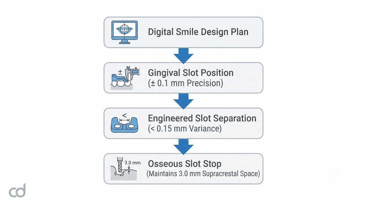
- Slot Separation Accuracy: The distance between the gingival and osseous rails is maintained within <0.15 mm, ensuring that the 3.0 mm supracrestal dimension is preserved across central incisors, lateral incisors, and canines.
- Gingival Slot Depth Tolerance: Soft-tissue margins are controlled within 0.1 mm, preventing over-resection that could expose sensitive root surfaces or violate interdental papillae.
- Rigid Seating Fit: Tooth-borne internal surfaces maintain seating tolerances below 0.05 mm, preventing rotational movement during instrumentation.
Section 6: Clinical Benefits & Practice Impact

Patient-Level Clinical Benefits
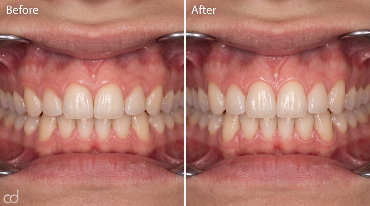
- Predictable Aesthetic Harmony: Gingival zeniths and exposed tooth proportions match the approved digital preview, giving patients confidence in their aesthetic treatment.
- Protection from Tissue Rebound: Controlled bone reduction maintains the natural supracrestal attachment apparatus, preventing late-stage tissue creep and margin relapse.
- Reduced Postoperative Morbidity: Guided micro-incisions and targeted osteoplasty minimize unnecessary tissue trauma, resulting in less swelling, faster re-epithelialization, and less post-op discomfort.
Practice-Level Operational Advantages
- Routine Adoption of Guided Gingivectomies: The system standardizes a procedure that is often operator-dependent, enabling restorative teams to execute crown lengthening with high repeatability.
- Workflow Cost-Efficiency: Combining soft-tissue resection and osseous reduction into a single template run shortens operatory time and eliminates separate corrective touch-up appointments.
- Reliable Restorative Timelines: Stable tissue margins heal predictably, allowing restorative teams to scan and prepare final veneers or crowns at 6 weeks post-op without fear of late-stage gingival migration.
- Reduced Intraoperative Stress: Pre-planned cutting rails eliminate real-time freehand calculations, allowing the surgical team to focus on clean execution and flap management.
The Modern Standard for Anterior Periodontal Aesthetics
Freehand aesthetic crown lengthening relies heavily on subjective visual estimates that can lead to uneven margins and biological width violations. The Curedent dual-slot guide system replaces freehand variability with digital predictability. By integrating smile design parameters into a rigid, dual-rail template, clinicians can achieve stable, symmetrical gingival margins with high confidence.
Section 7: Ordering, FAQ & Call to Action
Simple Three-Step Ordering Workflow
Integrating the Curedent Gum Reduction Guide into your practice follows an efficient three-step process:
- Upload Case Records: Access the Curedent portal and upload the patient’s optical intraoral scans (maxillary and mandibular STL/PLY) alongside high-resolution retracted and dynamic facial photographs.
- Review Digital Design: Curedent engineers integrate the facial landmarks, design the target tooth proportions, and position the dual-slot guide rails. Review the 3D smile proposal on your web browser, request adjustments if desired, and confirm approval.
- Receive Surgical Kit: Curedent 3D-prints the guide at 50-micron resolution, inspects slot tolerances, packages the appliance in protective, sterilization-ready casing, and dispatches the kit directly to your clinic.
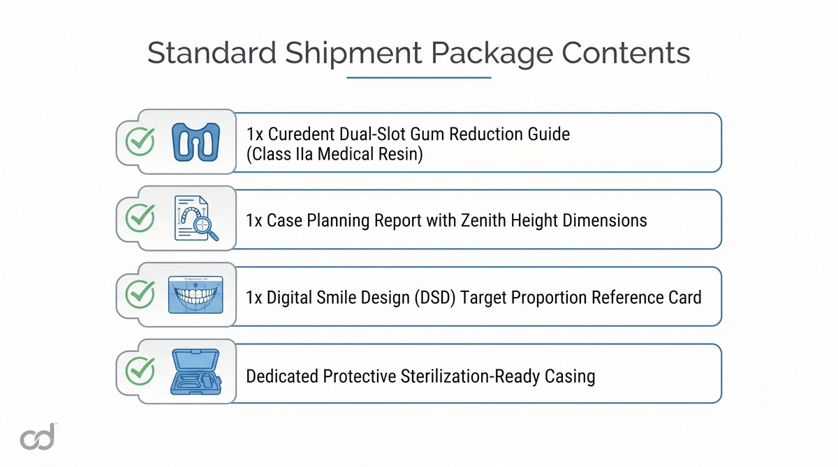
Standard Shipment Package Contents:
———————————–
[✓] 1x Curedent Dual-Slot Gum Reduction Guide (Class IIa Medical Resin)
[✓] 1x Case Planning Report with Zenith Height Dimensions
[✓] 1x Digital Smile Design (DSD) Target Proportion Reference Card
[✓] Dedicated Protective Sterilization-Ready Casing
Frequently Asked Questions
Do I need to submit a completed Digital Smile Design (DSD) file?
No. You can upload raw intraoral scans and 2D facial photographs; Curedent digital technicians will generate the 3D smile design and position the dual-slot rails for your review. If you already have an in-house DSD setup, simply upload the project files.
Can this guide be used with dental lasers as well as standard scalpels?
Yes. The coronal gingival slot accommodates both standard micro-scalpel blades (e.g., #15C, Beaver blades) and diode or erbium laser tips up to 0.8 mm in diameter.
What should I do if the guide does not seat passively on the incisal edges?
Inspect the incisal windows. If an air gap shows, check for newly placed composite restorations, interproximal calculus, or unblocked undercuts. Relieve minor internal acrylic contacts along non-functional line angles using a fine acrylic bur until the guide seats passively.
Is the Curedent guide resin certified for intraoral surgical contact?
Yes. Every guide is 3D-printed from certified Class IIa biocompatible surgical photopolymer, ensuring it is non-toxic, non-irritating, and dimensionally stable during surgery.
How does the guide protect against biological width violations?
The appliance mechanically controls bone reduction: the apical osseous slot is locked 3.0 mm apical to the coronal gingival slot, ensuring that crestal ostectomy preserves the natural 2.04 mm supracrestal tissue attachment plus physiological sulcus depth.
Suggested Clinical Solutions
- Transfer complex full-arch osteotomies with the Curedent Stackable Surgery Guide.
- Ensure accurate single and multi-unit implant placements using the Curedent Single Implant Surgical Guide.
- Protect newly contoured restorations and maintain stability with Curedent Clear Retainers.