Section 1: Product Introduction & Clinical Purpose
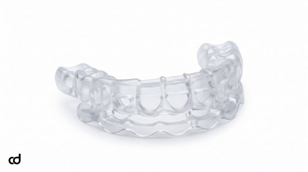
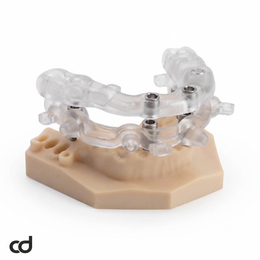
The Curedent Orthognathic Surgery Surgical Guide System is a patient-specific, CAD/CAM-manufactured intraoperative positioning appliance set comprising high-precision intermediate wafers and final occlusal splints. Developed specifically for oral and maxillofacial surgeons, surgical orthodontists, and virtual planning teams, this system provides the physical bridge between digital skeletal simulations and operative theater reality.

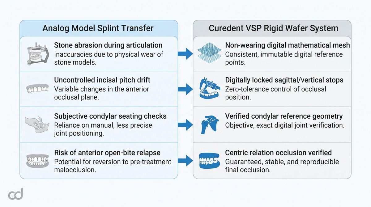
Translating virtual surgical planning (VSP) into intraoperative execution has historically relied on manual stone-cast model surgery, facebow transfers, and hand-cured acrylic splints. These analog techniques introduce cumulative mechanical discrepancies at every laboratory bench step, leading to unpredictable intermediate positions, yaw/pitch errors, and uncorrected facial asymmetries. The Curedent orthognathic system eliminates these transfer variances by converting voxel-accurate digital simulations directly into rigid, 3D-printed positioning wafers that lock osteotomized bony segments into exact spatial coordinates.
Key Clinical Advantages
- High-Fidelity Virtual Transfer: Translates computer-planned multi-planar translations, advancements, and roll/pitch/yaw corrections directly to the operatory.
- Rigid Spatial Stabilization: Maintains intersegmental positioning against soft-tissue resistance and muscle pull during preliminary fixation.
- Streamlined Multi-Piece Tracking: Accommodates segmentalized maxillary designs and asymmetric bilateral ramus shifts without intraoperative rocking.
- Maximized Intraoperative Efficiency: Eliminates chairside acrylic grinding and re-indexing, shortening general anesthesia duration.
 .
Section 2: Product Anatomy & Physical Construction
Medical-Grade Rigid Polymer Construction
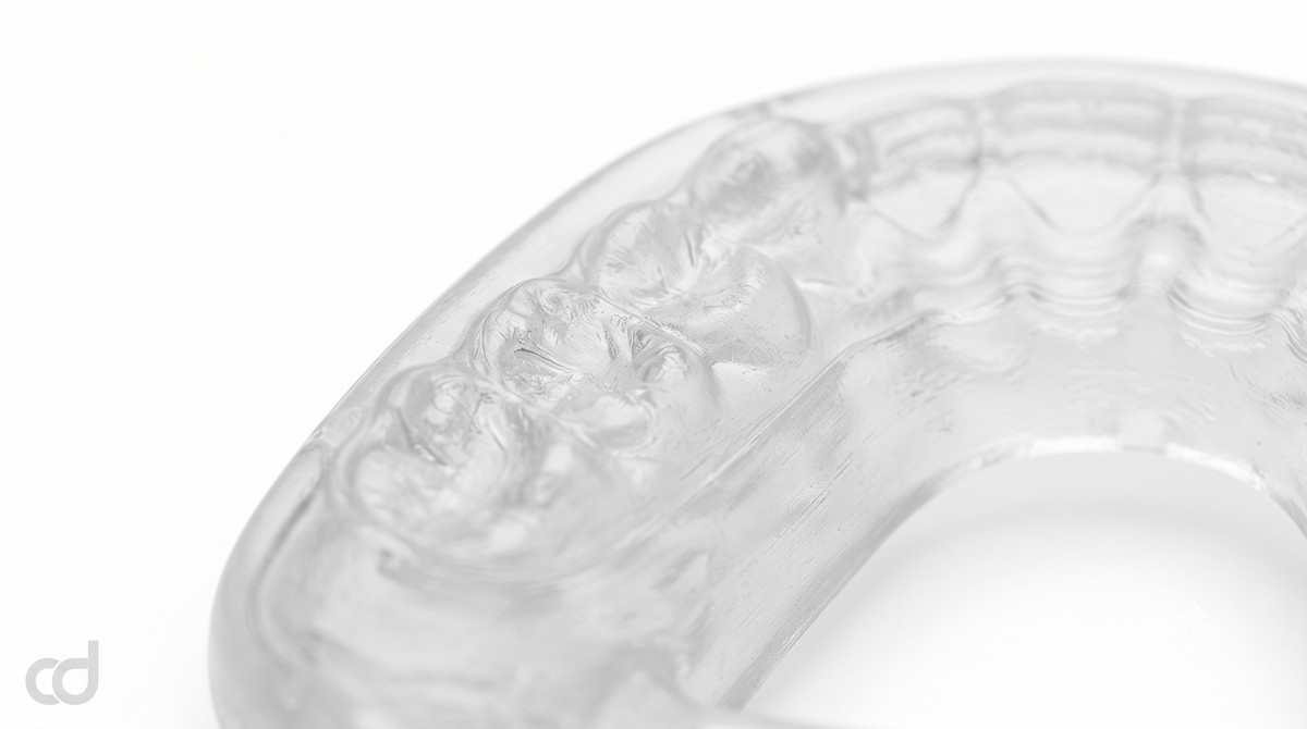
The Curedent orthognathic appliance set is manufactured from an FDA-compliant, Class IIa certified biocompatible surgical photopolymer. The material features high flexural strength, high modulus of elasticity, and minimal moisture absorption in the presence of blood, irrigation fluids, and saliva. This dimensional resilience ensures the wafer will not warp, deform, or compress under high mechanical loads during surgical osteotomy manipulation.
High-Resolution Additive Manufacturing
Using calibrated Digital Light Processing (DLP) and Stereolithography (SLA) printing systems running at ~50-micron layer resolution, every wafer achieves precise registration against brackets, archwires, and enamel surfaces. The fine z-axis resolution eliminates layer stepping on functional inclines, ensuring that bracket wings and molar tubes do not bind against the appliance during insertion.
Structural Anatomy of the Intermediate Wafer
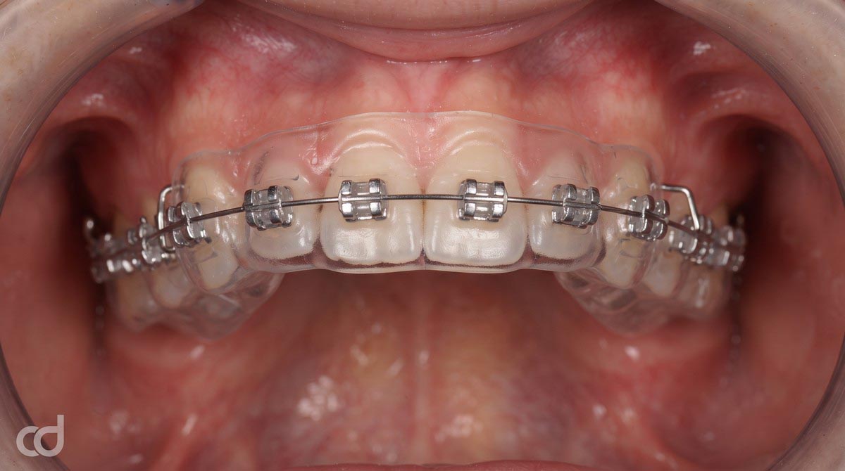
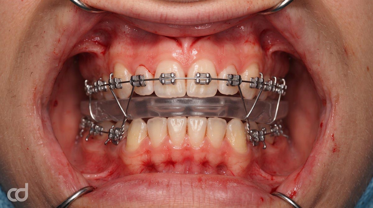
The intermediate surgical wafer indexes the relationship between the mobilized maxilla (or mandible, depending on sequence) and the unmoved, intact opposing arch. It features:
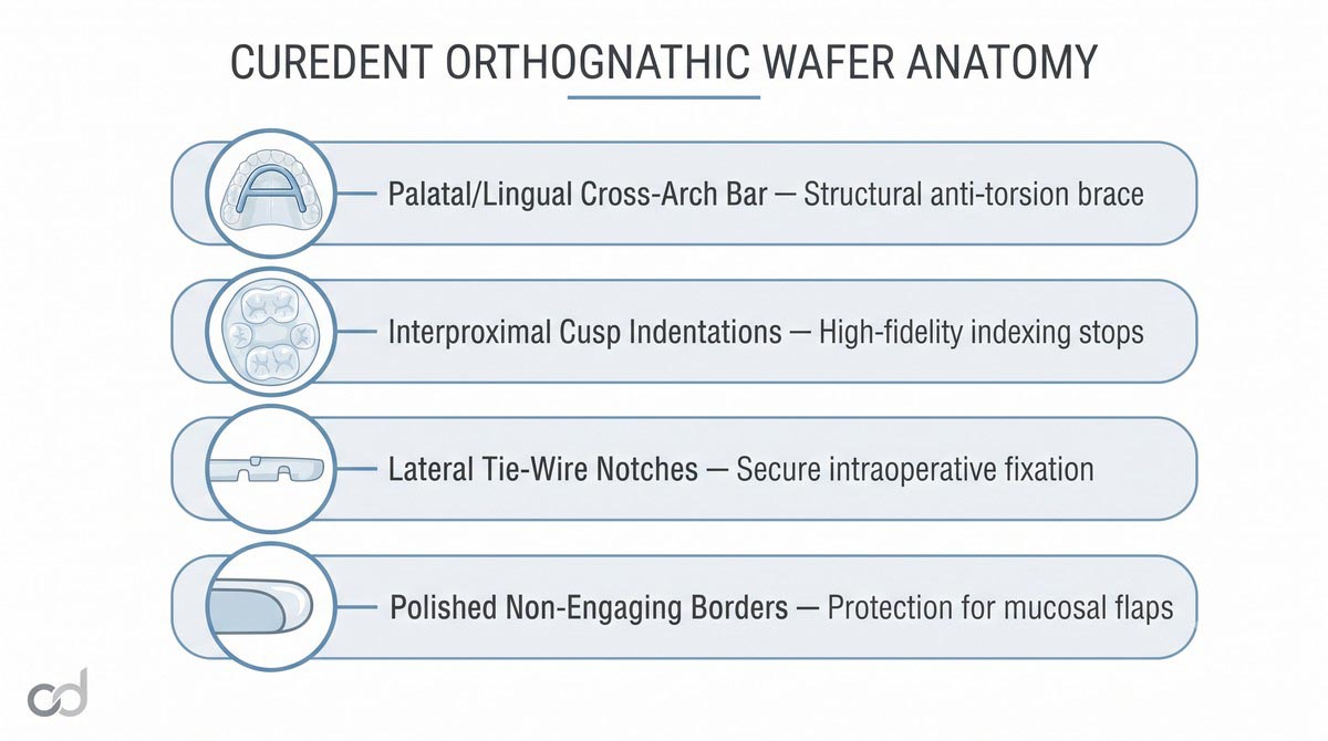
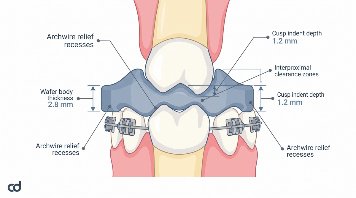
- Deep Cuspal Indexing: Cusp indentations are digitally contoured to a depth of 1.0–1.5 mm to provide positive stops without trapping bracket ties.
- Cross-Arch Reinforcing Struts: Palatal or lingual connecting bars reinforce structural rigidity, preventing cross-arch flexure when stabilizing the mobilized jaw.
- Fixation Wire Notches: Recessed lateral notches allow direct stabilization to orthodontic archwires using 0.010-inch stainless steel ligature wires.
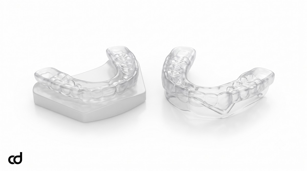
Structural Anatomy of the Final Orthognathic Splint
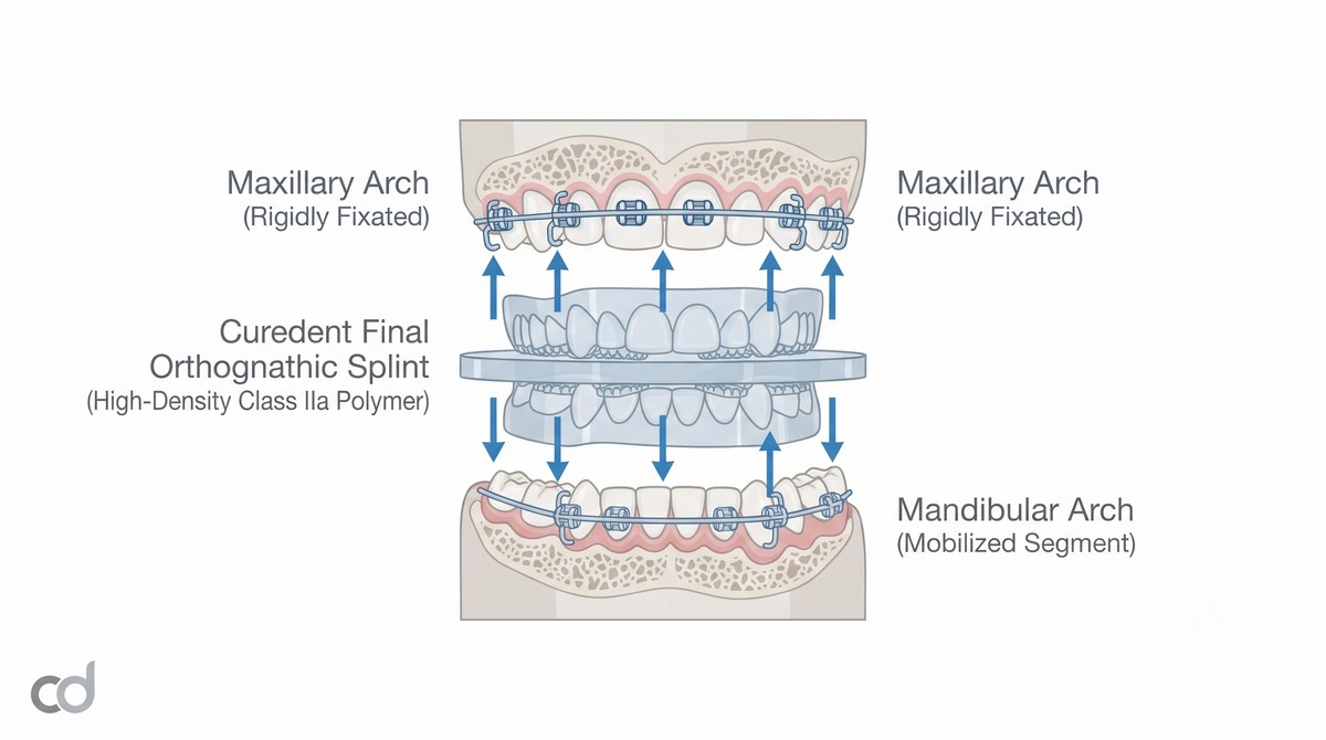
The final splint establishes definitive occlusal interdigitation between the newly osteotomized maxilla and mandible. Designed to seat passively across both arches, it provides verified stops for all load-bearing cusps, ensuring the condyles remain seated within the glenoid fossae while internal rigid fixation plates and screws are applied.
Mechanical Integrity of the 3D Printed Rigid Wafer
A critical functional requirement of any 3D printed rigid wafer is dimensional stability under operative loads. Hand-mixed analog cold-cure acrylic splints often exhibit internal porosity, uneven polymerization shrinkage, and elastic flexure when exposed to manual seating forces. In contrast, Curedent additive photopolymers undergo controlled post-print ultraviolet thermal curing, producing an isotropic, high-density polymer network that resists cross-arch deformation when the surgeon manipulates the mobilized skeletal segments into position.

Section 3: Indications & Clinical Applications
Primary Clinical Indications
The Curedent Orthognathic Surgery Surgical Guide System is indicated across the spectrum of complex corrective jaw procedures within modern maxillofacial surgery:
- Skeletal Class III Discrepancies: Severe mandibular prognathism, midface hypoplasia, or combined discrepancies requiring maxillary advancement, maxillary downgrafting, and mandibular setback.
- Skeletal Class II Discrepancies: Severe retrognathia and mandibular hypoplasia requiring large mandibular advancements, often combined with counter-clockwise pitch rotation of the maxillomandibular complex.
- Facial Asymmetry and Canting: Asymmetric occlusal plane cants, yaw deviations, and hemimandibular hyperplasia requiring differential multi-planar adjustments.
- Multi-Piece Segmental Maxillary Osteotomies: 2-piece or 3-piece Le Fort I procedures addressing transverse arch collapse, curve of Spee leveling, and tooth-size discrepancies.
- Vertical Maxillary Deformities: Vertical maxillary excess (gummy smile) requiring superior impaction, or vertical deficiency requiring interpositional bone grafting.
Orthognathic Surgical Assessment Checklist:
[✓] Decompensation complete: Dental arches leveled, aligned, and coordinated
[✓] Orthodontic appliances stable: Heavy passive rectangular archwires secured
[✓] Imaging current: High-resolution CBCT capturing condyles to menton (< 4 weeks old)
[✓] Virtual setup validated: Overbite, overjet, midline, and soft-tissue profile confirmed
[✗] Active dental movements ongoing without stable surgical archwires (Contraindicated)
Case Selection Criteria
- Completed Presurgical Orthodontic Decompensation: Teeth must be uprighted over basal bone with arch coordination established, eliminating dental compensations that could disguise skeletal discrepancies.
- Rigid Surgical Archwires: Patient dentition must carry passive, full-dimension rectangular archwires (e.g., 0.0190.025-inch stainless steel) with soldered or crimped surgical hooks to secure the wafer and support intraoperative fixation.
- Accurate Preoperative Imaging: Diagnostic records must include a volumetric CBCT with condyles fully seated, free from patient motion artifacts, along with clear optical scans of the dentition.
Guided Wafer Accuracy in Skeletal Class III Correction
In cases of skeletal Class III correction, managing maxillary advancement while setting the mandible back requires precision across all three spatial dimensions. Freehand repositioning or analog stone-model splints carry high rates of rotational yaw and pitch error:
Section 4: Clinical Workflow & Surgical Execution

Step 1: Comprehensive Data Acquisition
Acquire a full-head or maxillofacial CBCT scan with the patient’s condyles properly seated in centric relation, relaxed musculature, and stable lip posture. Capture full-arch optical intraoral scans of both maxilla and mandible, detailing occlusal anatomy, bracket geometry, and surgical hooks.
Step 2: Virtual Surgical Planning (VSP)
Upload datasets to the Curedent digital engineering portal. In collaboration with the surgical team, Curedent engineers segment the skeletal framework, define osteotomy cut lines, calculate exact translation vectors (roll, pitch, yaw, advancement, impaction), and generate digital models for the intermediate wafer and final splint.
Step 3: Unboxing and Pre-Surgical Verification
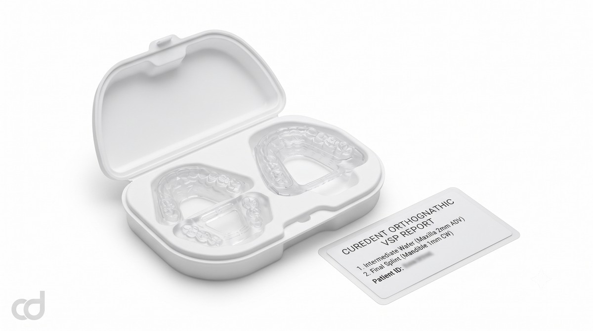
The guide set arrives in validated, sterilization-ready packaging. Prior to scrub-in, verify that the set contains:
- 1x Intermediate Surgical Wafer (labeled with designated sequence)
- 1x Final Orthognathic Splint
- Comprehensive VSP translation report card detailing millimeter and angular movements
Step 4: Intermediate Wafer Execution (Maxilla-First or Mandible-First)
In a standard maxilla-first protocol:
- Perform downfracture during the Le Fort I osteotomy, mobilizing the maxilla completely from the pterygoid plates and nasal septum.
- Seat the intermediate surgical wafer onto the intact, unoperated mandibular arch.
- Bring the mobilized maxilla down into the superior indexing surface of the intermediate wafer.
- Apply temporary intermaxillary fixation wires to hold the maxilla, wafer, and mandible as a single unit. Ensure the condyles remain fully seated in the glenoid fossae.
- Apply titanium miniplates and monocortical screws along the pyriform apertures and zygomaticomaxillary buttresses to achieve rigid internal fixation.
- Release the temporary fixation wires and remove the intermediate wafer.
Step 5: Second Jaw Mobilization and Final Splint Placement
- Perform the bilateral sagittal split osteotomy (BSSRO) on the mandible, mobilizing the distal tooth-bearing segment from the proximal condylar-bearing segments.
- Seat the final orthognathic splint between the maxillary and mandibular dentition.
- Establish definitive intermaxillary fixation using wire ligatures or elastics across surgical archwire hooks.
- Position the proximal ramus segments to ensure the condyles seat properly within the glenoid fossae, preventing posterior or lateral displacement.
- Apply rigid internal fixation (bicortical position screws or monocortical plate systems) across the mandibular osteotomy sites.
Step 6: Final Verification and Release
Release the temporary intermaxillary fixation wires. Rotate the mandible open and closed on its terminal hinge axis to confirm smooth, reproducible interdigitation into the splint without slide, muscular resistance, or occlusal interference. The splint may be wired to the maxillary arch for light post-surgical guiding elastics.
Surgical Safety: Seating Verification and Condylar Alignment
Always confirm that bracket wings and ligature wires seat cleanly within the pre-planned relief grooves. If a bracket contacts the acrylic border before cusps reach full depth, the osteotomy position will be displaced. Prior to final fixation, the surgeon must gently palpate and seat the condyle into the anterior-superior position within the glenoid fossa. Securing plates while the condyle is distracted or torqued introduces significant risk of postoperative skeletal relapse, malocclusion, and temporomandibular joint dysfunction.

Section 5: Technical Specifications & Accuracy Standards
Certified Technical Specifications
The Curedent Orthognathic Surgery Guide System is manufactured to strict biomedical standards to ensure dependable translation from digital plan to surgical execution.
| Parameter | Specification | Clinical Relevance |
| Material | Class IIa biocompatible rigid surgical resin | Rigid structural stability; zero intraoral toxic leaching |
| Printing method | DLP / SLA stereolithography | Captures bracket clearances and fine cuspal indentations |
| Layer resolution | ~50 microns | Smooth seating surfaces without stepping artifacts |
| Product types | Intermediate surgical wafer + final orthognathic splint | Coordinates multi-jaw osteotomy transitions |
| Wafer thickness | 2.5–4.0 mm (case-dependent) | Balances interocclusal space against fracture resistance |
| Occlusal registration accuracy | < 0.1 mm | Preserves digital plan accuracy across all dimensions |
| Flexural rigidity | Engineered to withstand IMF wire tension | Prevents cross-arch deformation under heavy wire forces |
| Dimensional stability | Stable across steam sterilization cycle | Resists thermal distortion and volumetric shrinkage |
| Sterilization | Autoclavable / sterilization-ready delivery | Complies with sterile surgical operating room protocols |
| Compatibility | Compatible with standard IMF wiring systems | Fits standard archwires, surgical hooks, and screws |
Precision Performance Metrics
Physical wafer accuracy determines how faithfully the virtual plan transfers to the patient:

- Intermediate Surgical Wafer Fidelity: The intermediate surgical wafer maintains dimensional registration within <0.1 mm across the full arch, ensuring accurate sagittal and vertical translation of the initial osteotomized jaw.
- Final Orthognathic Splint Interdigitation: The final orthognathic splint seats with balanced bilateral contacts, ensuring passive seating over orthodontic hardware and establishing the planned class I canine and molar relationships.
- Thermal and Autoclave Stability: Formulated to withstand steam autoclave cycles without warping, shrinking, or undergoing plastic deformation, ensuring accurate fit on the operating table.
Section 6: Clinical Benefits & Practice Impact

Patient-Level Clinical Benefits
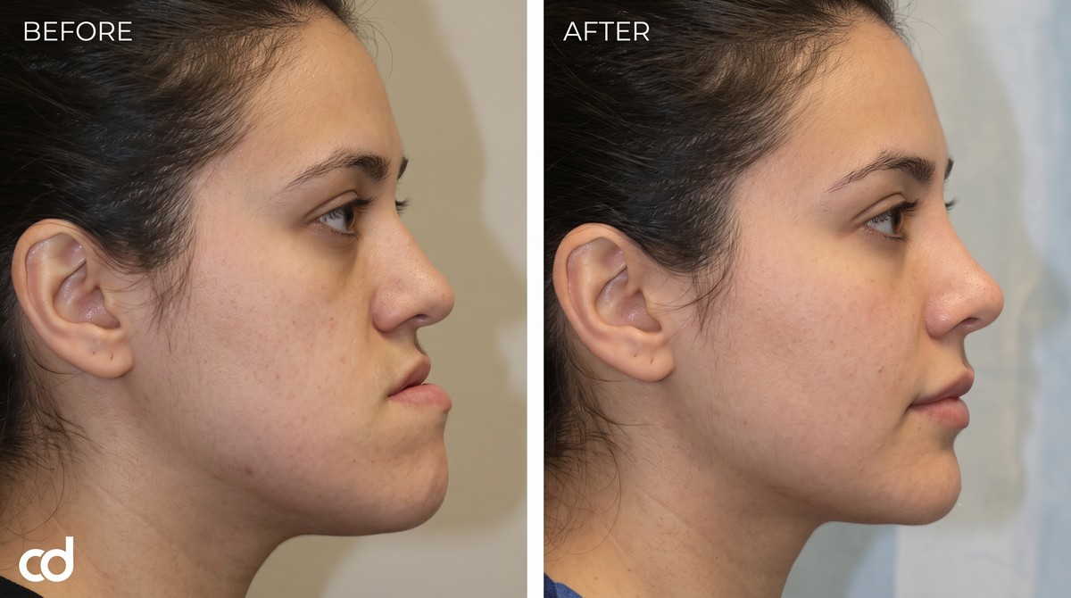
- Predictable Facial Symmetry and Aesthetics: Digital planning translates directly to the patient, ensuring planned chin projections, lip supports, and midline alignments match the final facial profile.
- Reduced General Anesthesia Time: Eliminating intraoperative splint adjustment and bite equilibration shortens overall operative time, lowering anesthetic exposure and recovery times.
- Lower Relapse and Revision Risks: Verified condylar seating during fixation minimizes internal stress on the temporomandibular joint, protecting against postoperative skeletal relapse.
Practice-Level Operational Advantages
- Routine Adoption of VSP Protocols: Removes the lab bottlenecks of stone-model surgery, facebow mountings, and manual acrylic fabrication, allowing surgical teams to manage complex deformities within a streamlined digital workflow.
- High Workflow Cost-Efficiency: Standardized digital workflows reduce intraoperative delays, eliminate consumable lab waste, and prevent costly secondary revision procedures.
- Reduced Intraoperative Stress: Pre-planned occlusal stops eliminate guesswork when adjusting bone plates, allowing surgeons to focus on osteotomy safety and soft-tissue closure.
- Resistance to Operative Loads: Engineered to withstand intermaxillary fixation (IMF) forces without cracking, flexing, or undergoing plastic deformation under stainless steel wire tension.
The Modern Standard for Corrective Jaw Surgery
Analog model surgery and hand-cured acrylic wafers are increasingly obsolete in modern orthognathic care. Additive CAD/CAM manufacturing, volumetric CBCT diagnostics, and virtual surgical planning have made digital wafer systems the gold standard. Curedent orthognathic wafers give surgical teams the structural rigidity and micro-meter precision needed to execute complex maxillofacial corrections with confidence.
Section 7: Ordering, FAQ & Call to Action
Simple Three-Step Ordering Workflow
Integrating the Curedent Orthognathic Surgery Guide System into your surgical planning workflow follows a straightforward sequence:
[Step 1: Upload Records] ──► [Step 2: VSP Verification] ──► [Step 3: Sterile Delivery]
CBCT + Optical Scan Files Interactive Translation Plan Pre-Sterilized Wafer Set
- Upload Case Records: Access the Curedent portal and upload the patient’s CBCT DICOM files, dental optical scans (STL/PLY), and presurgical orthodontic photos.
- Interactive VSP Review: Collaborate with Curedent biomedical engineers to review skeletal osteotomies, approve translational movements, confirm occlusal clearances, and finalize intermediate and final splint designs.
- Receive Surgical Wafers: Curedent fabricates the wafer set at 50-micron resolution, confirms dimensional accuracy, packages the components in protective, sterilization-ready casing, and dispatches the kit directly to your facility.
Standard Shipment Package Contents:

Frequently Asked Questions
Do I need to provide a completed VSP file, or can Curedent perform the planning?
Curedent supports both workflows: you can upload your completed VSP design files for immediate printing, or provide raw scans and work directly with our engineering team to plan the skeletal osteotomies and splints.
Can I use standard fixation screws and plates with these wafers?
Yes. Curedent wafers index the dentition and archwires, leaving buccal bone surfaces and osteotomy cut margins clear for standard miniplate and screw fixation systems.
What resin material is used for the wafers?
All appliances are printed using certified Class IIa biocompatible surgical resins that meet international standards for intraoperative mucosal and bone contact.
Can the 3D-printed wafer withstand heavy IMF wire tension?
Yes. The Class IIa resin is formulated for high flexural strength and modulus of elasticity, preventing deformation or cracking when torqued down with 0.010-inch stainless steel ligature wires.
How does the final splint differ from the intermediate wafer?
The intermediate wafer indexes the mobilized first jaw against the stationary opposing arch to set the intermediate position. The final splint indexes the completed, definitive occlusion between both mobilized jaws for final plating and postoperative stabilization.
Suggested Clinical Solutions
- Manage full-arch implant restorations with the Curedent Stackable Surgery Guide.
- Ensure accurate single and multi-site osteotomies with the Curedent Single Implant Surgical Guide.
- Protect teeth and stabilize postoperative splints with Curedent Clear Retainers.